Your doctor can use the images from an echocardiogram to identify heart disease. Depending on what information your doctor needs, you may have one of several types of echocardiograms. Each type of echocardiogram has few, if any, risks involved.
Your doctor may suggest an echocardiogram if he or she suspects problems with the valves or chambers of your heart or if heart problems are the cause of symptoms such as shortness of breath or chest pain. An echocardiogram can also be used to detect congenital heart defects in unborn babies (fetal echocardiogram).
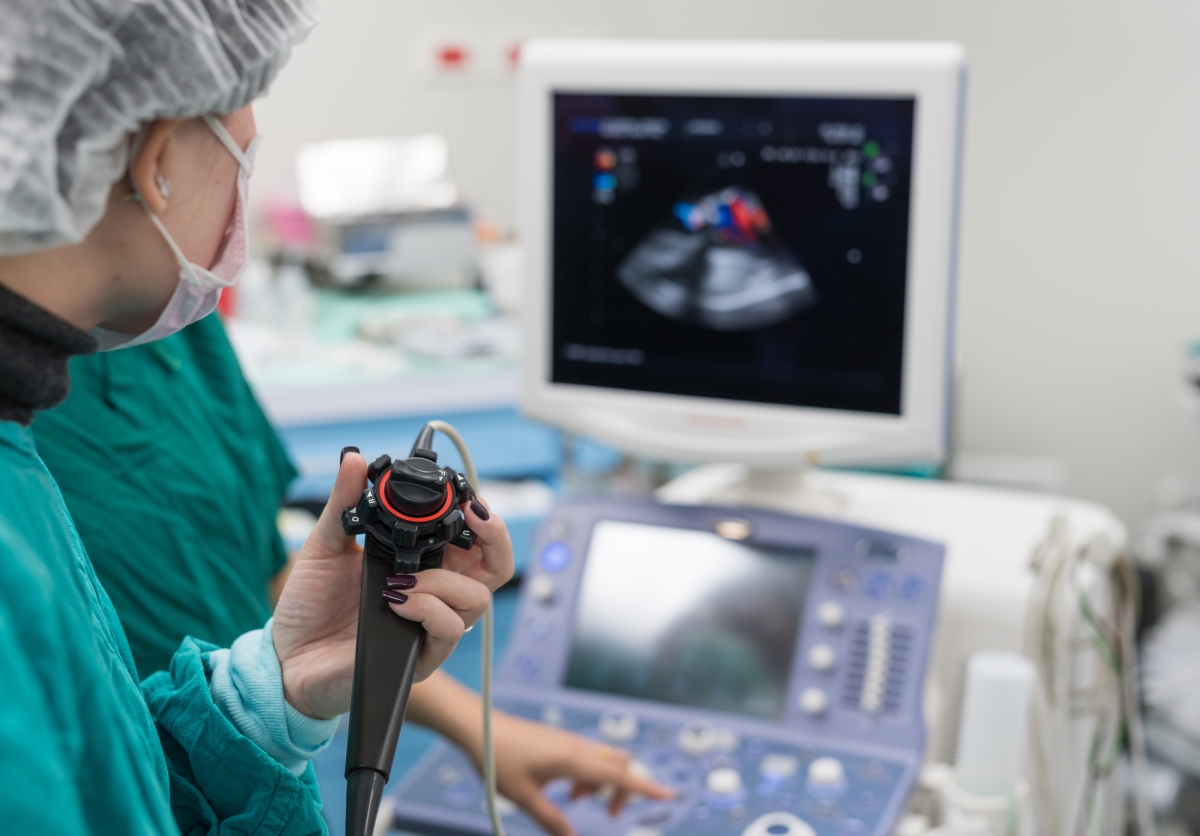
This is a standard, noninvasive echocardiogram. A technician (sonographer) spreads gel on your chest and then presses a device known as a transducer firmly against your skin, aiming an ultrasound beam through your chest to your heart. The transducer records the sound wave echoes from your heart. A computer converts the echoes into moving images on a monitor. If your lungs or ribs block the view, you may need a small amount of liquid (contrast agent) injected through an intravenous line (IV) that will make your heart’s structures show up more clearly on a monitor, improving the images.
If it’s difficult to get a clear picture of your heart with a standard echocardiogram or if there is a reason to see the heart and valves in more detail, your doctor may recommend a transesophageal echocardiogram. In this procedure, a flexible tube containing a transducer is guided down your throat and into your esophagus, which connects your mouth to your stomach. From there, the transducer can be positioned to obtain more-detailed images of your heart. Your throat will be numbed, and you’ll have medications to help you relax during a transesophageal echocardiogram.
When sound waves bounce off blood cells moving through your heart and blood vessels, they change pitch. These changes (Doppler signals) can help your doctor measure the speed and direction of the blood flow in your heart. Doppler techniques are used in most transthoracic and transesophageal echocardiograms, and they can be used to check blood flow problems and blood pressures in the arteries of your heart that traditional ultrasound might not detect. Sometimes the blood flow shown on the monitor is colorized to help your doctor pinpoint any problems..
Some heart problems — particularly those involving the coronary arteries that supply blood to your heart muscle — occur only during physical activity. For a stress echocardiogram, ultrasound images of your heart are taken before and immediately after walking on a treadmill or riding a stationary bike. If you’re unable to exercise, you may get an injection of a medication to make your heart pump as hard as if you were exercising.
There are no risks involved in a standard transthoracic echocardiogram. You may feel some discomfort similar to pulling off an adhesive bandage when the technician removes the electrodes placed on your chest during the procedure. If you have a transesophageal echocardiogram, your throat may be sore for a few hours afterward. Rarely, the tube may scrape the inside of your throat. Your oxygen level will be monitored during the exam to check for any breathing problems caused by sedation medication. During a stress echocardiogram, exercise or medication — not the echocardiogram itself — may temporarily cause an irregular heartbeat. Serious complications, such as a heart attack, are rare.
Your doctor will look for healthy heart valves and chambers, as well as normal heartbeats. Information from the echocardiogram may show:
Weakened or damaged heart valves, high blood pressure, or other diseases can cause the chambers of your heart to enlarge or the walls of your heart to be abnormally thickened. Your doctor can use an echocardiogram to evaluate the need for treatment or monitor treatment effectiveness.
An echocardiogram can help your doctor determine your heart’s pumping strength. Specific measurements may include the percentage of blood that’s pumped out of a filled ventricle with each heartbeat (ejection fraction) or the volume of blood pumped by the heart in one minute (cardiac output). If your heart isn’t pumping enough blood to meet your body’s needs, this could result in heart failure.
During an echocardiogram, your doctor can determine whether all parts of the heart wall are contributing normally to your heart’s pumping activity. Parts that move weakly may have been damaged during a heart attack or be receiving too little oxygen. This may indicate coronary artery disease or various other conditions..
An echocardiogram shows how your heart valves move as your heart beats. Your doctor can determine if the valves open wide enough for adequate blood flow or close fully to prevent blood leakage.
Many heart defects can be detected with an echocardiogram, including problems with the heart chambers, abnormal connections between the heart and major blood vessels, and complex heart defects that are present at birth. Echocardiograms can even be used to monitor a baby’s heart development before birth.